Articles
Advances in Medical Imaging Technology

Medical imaging - an application of biomedical optics in the field of healthcare - is the graphic visualization of the body for use in diagnosis, treatment and monitoring of medical conditions.
Imaging technologies play a vital role in making medical procedures more scientific as they help confirm or reject the diagnostician’s or physician’s initial hypothesis about the patient’s ailment.
It adds a whole new dimension to medical diagnosis applications, making the process faster and more accurate, and helps predict or discover previously undetected, intricate rare conditions.
The drive for more emergent bio-optical devices has lead to the foundation of the field of diagnostic medicine in which, certain compounds, which are engineered to react only to specific conditions, are revealed only by specific electromagnetic (EM) wavelengths. This helps in diagnosis rare diseases and other specialty conditions.
And not only diagnosis and treatment, optics are increasingly being used in surgery procedures, or so called image-guided surgery.
Scientists are innovating newer imaging modalities by harnessing previously untapped wavelengths of the EM spectrum. Their quantum leaps in understanding biomedical optics has lead to more perspicuity on ailments such as certain types of cancer, Parkinson’s disease and Alzheimer’s disease.
The medical imaging market has been growing at an explosive rate and it is necessary that medical professionals know about the future of the field and the emergent, upcoming imaging devices and technologies.
1. Hyperspectral Imaging
Hyperspectral imaging (HSI) lubricates digital imaging with spectroscopy to obtain the entire spectral data of EM waves for each individual pixel.
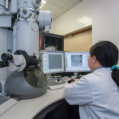
HSI finds applications in a many fields, including surveillance, agriculture, eye care, mineralogy and astronomy. Some popular examples where this is used is in electron microscopes.
Due to its ability to give both spatial and spectral data non-invasively in three dimensions, biomedical engineers are incorporating HSI in imaging cancerous tissues, pathology and tissue morphology, physiology and composition, as well as guiding surgeries.
HSI works on the observation that biological tissues, when incident by light, undergoes erratic scattering from the inhomogeneity of biological structures, and absorption primarily in haemoglobin, melanin, and water as it propagates through the tissue.
Scientists logically conclude that this would also change the absorption, fluorescence, and scattering characteristics of the tissue as the disease progresses.
This scattered, fluorescent and reflected light from the tissue captured carries quantitative diagnostic information about tissue pathology.
HSI works on the mathematical model of a virtual 3D dataset cube with two spatial dimensions and one spectral. The camera comprises of a light source, wavelength dispersion devices, and area detectors.
It acquires data using four main acquisition techniques - spatial scanning, spectral scanning, non-scanning and spatio-spectral scanning. HSI could help reduce surgery complications and associated liability.
2. Mega Microchip
Conceptualized in 2011, the DynAMITe microchip, which is still in its prototype phase, takes high-resolution images of the effects of radiation on cancerous tumours. Its unique identification is its large size.
The device itself can withstand radiation levels above the maximum limits in radiation therapy procedures.
Based on Complementary Metal Oxide Semiconductor (CMOS) technology, the microchip is an 0.18-µm active pixel sensor with a 1280×1280 pixels per 100-µm pitch and 2560×2560 pixels per 50-µm pitch.
It can be operated at rates of up to 90 fps. Arrays of 2×2 devices can be assembled with minimal border loss, providing an imaging area greater than 25 cm2.
DynAMITe essentially replaces the conventional amorphous silicon panel technology and has larger pixels than those found in consumer digital cameras or mobile phones. It can be used in mammography and radiotherapy as it will withstand exposure to very high levels of x-ray and other radiation.
3. Holey Metamaterial
Although reliable, a major drawback ultrasound imaging faces is its inability to obtain high-resolution, detailed images.
To compensate for this shortcoming, a team of scientists at UC Berkeley (USA) and Universidad Autonoma de Madrid (Spain) invented a 3D metamaterial to achieve “deep sub-wavelength imaging”.
This metamaterial will likely enhance the image resolution by a factor of 50.
The material can be incorporated into already functioning ultrasound probes to capture high-res medical images.
The metal metamaterial is composed of 1,600 hollow copper tubes measuring roughly 1 mm in diameter; the tubes are bundled into a 6-in bar featuring a 2.5-in2 cross-section.
The working principle underlying this material is as follows: in acoustic imaging, the size of the smallest feature is limited by diffraction, which restricts the resolution of the object to the scale of the wavelength of sound waves.
But the diffraction limit is valid only in the “far field”. Thus, higher-resolution imaging can be achieved by capturing the information contained within the quickly-fading waves of the “near field”.
This wave in the near-field carries a lot of information about the deep sub-wavelength features of this imaging object. Transferring this information to a near field will help reduce the frequency while increasing the resolution. Therefore higher frequencies can be achieved at much lower-frequency ultrasonic waves.
The lower the frequency, the better the penetration and the stronger the signal. As low as 1/50th of a wavelength can be seen in high resolution.
4. Palm-Size Magnetic Resonance Imaging - Superconducting Magnet System
Most MRIs these days use superconductors. However, their size hasn’t reduced and still use liquid helium to cool the superconducting wires. But there is still scope for further innovations which might lead to more mobile imaging applications.
The breakthrough was made back in 2011, when scientists in Tokyo, Japan, created the world’s smallest superconducting magnet that could fit in the palm. These were modelled after Japan’s levitating train Maglev.
This technology was envisioned to be resin-impregnated in order to avoid the need for liquid helium. Instead, according to the scientists, a small quantity of liquid nitrogen is enough to keep it cooled at superconductor levels.
This MRI is being designed to be used on the go.
However, modern MRIs still use bulky superconductors for magnets.


